SEM, FEI Quanta 600 FEG
FEI Quanta 600 FEG
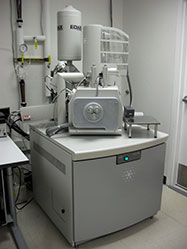
The Quanta 600 is a State-of-the-Art scanning electron microscope (SEM) with a high-resolution field-emission source. The system can operate in high-vacuum, low-vacuum, and true environmental (water vapor ambient) modes. The electron detectors included with this system are an Everhart-Thornley secondary electron detector, a large-field detector, a gaseous secondary electron detector, and a solid-state backscattered electron detector. A peltier cooled sample holder is available to aid in imaging biological and "wet" specimens. Analytical options will include an EDX system for compositional analysis, and an electron backscattered diffraction (EBSD) system for phase ID and to measure crystal orientation. This instrument was purchased through exceptionally generous support from the Office of the Vice-President for Research, with the EBSD and complementary XRF system provided through the generous donation of George and Linda Caine.
Click here to view sample images!
For Appointments for SEM with operator assistance contact:
- Prof. Ian Harvey - irharvey@eng.utah.edu
- Dr. Brian Van Devener - bvandev@chem.utah.edu
- Dr. Paulo Perez - jperez@eng.utah.edu
Professional Technician time is available by appointment for SEM assistance.
Student Technicians:
- Sadee Hansen
- Nick Peterson
- Trevor Donney
Techniques Available:
Electron Microscopy
- Schottky field-emission source (thermally assisted)
- Hi-vac, low-vac, and ESEM modes
- ESEM enables hi-resolution topographical imaging in water vapor ambient
- Good for delicate samples otherwise damaged by vacuum
- Secondary and backscattered detectors for all pressure ranges
- Specified spot-size of < 5 nm in high-vacuum mode
- Large stage can mount and manipulate 6" Si wafers
- 5 axis-precision motorized stage
- Landing energy control system
EDX
- EDAX X-ray detector
- Detects B through U
- Detection limits ~ 0.1 to 1% atomic
- Element Mapping capability, with sub-micron resolution (specimen dependent)
EBSD
- Electron backscattered diffraction
- determines crystal structure of surface crystallites (phase identification)
- measures crystal orientation of surface crystallites (grain texture)
Sample Control and Preparation
- Large stage and chamber can hold and manipulate a 6" Si wafer
- 5 axis precision stage with compucentric rotation
- Peltier stage for sample cooling (biological and "wet" specimens)
- accelerating voltage control through deceleration, to 50 V landing energy
- A large variety of sample holders available
- Carbon tape, DAG, etc will be available to mount samples
Documentation
Downloads
- Casino (Simulation of Electron Trajectory in Solids)
Last Updated: November 7, 2011